Original Article - (2021) Volume 16, Issue 4
Transvaginal Power Doppler Sonography as an Adjunct Tool to Histopathology in Detection of Endometrial Changes in Women with Postmenopausal Bleeding
Sally AR Kotb1*, Nahla M Awad2 and Amal S Zaghlaul1Received: 29-Nov-2021 Published: 24-Dec-2021
Abstract
Background: Postmenopausal bleeding (PMB) represents a practical challenge due to the increased risk of endometrial carcinoma which requires efficient evaluation for optimum management. The use of the three vascular patterns that could be depicted by transvaginal Power Doppler sonography (TVPD) is useful to discriminate endometrial carcinoma from other benign conditions. The aim of this study was to evaluate TVPD in discrimination between benign and malignant endometrial lesions in PMB women compared to the histopathologic diagnosis.
Methods: Eligible 180 patients complaining of PMB were involved in the study, they were examined by Gray scale transvaginal sonography to assess the endometrium, TVPD scanning of the endometrial and endometrial myometrial interface vessels (Spiral arteries), followed by pulsed Doppler velocimetry and lastly endometrial biopsy for histopathological diagnosis.
Results: The endometrial characteristics and the different vascular patterns depicted by TVPD showed a good diagnostic ability in depicting endometrial malignancy and differentiate it from benign endometrium:16 cases (8.9%) showed multiple branching vessels pattern (pattern A) strongly correlated with endometrial cancer, 31 cases (17.2%) showed single non branching vessel (pattern B) suggesting polyp, 61 cases (33.9%) showed scattered vessel pattern (pattern C) suggestive of hyperplasia, and no power Doppler signals were detected in 72 cases (40%) diagnosed as atrophy. Atrophy has been found to be the commonest pathological lesions as a cause of PMB. Spiral arteries pulsatility index and resistance index were measured and were significantly lower in malignant endometrium compared to other histopathological benign endometrial pathology p <0.001.
Conclusion: Power Doppler ultrasound is a useful adjunct tool in diagnosis of endometrial pathology in cases with postmenopausal bleeding and shows good reliability in the discrimination between endometrial carcinoma from other benign endometrial conditions.
Keywords
Postmenopausal bleeding; Power Doppler; Transvaginal Power Doppler sonography
Introduction
Abnormal uterine bleeding in postmenopausal women is of special concern due to the probability of endometrial carcinoma [1]. For this age group, prompt evaluation is required to plan management. Several approaches were proved to be clinically useful in diagnosing endometrial pathology in cases presenting with postmenopausal bleeding (PMB). This includes transvaginal sonography (TVS), sonohysterography, hysteroscopy and endometrial sampling for pathological examination [2].
Power Doppler has been proposed as non-invasive screening method in PMB cases [3]. A good correlation has been found between the spiral artery flow velocity waveforms and the histopathological diagnosis in women with PMB [4]. Although some authors have argued that endometrial echo texture may help to differentiate carcinoma from polyps and hyperplasia [4], power Doppler imaging can depict the microvasculature patterns in the sub-endometrial zone. It assesses the vasculature of a lesion based on the amplitude of the Doppler signal which is superior to the Doppler frequency shift used in color Doppler. Power Doppler is also independent from the insonation angle and has high perception of low velocity blood vessels, depicting vasculature clearly and reliably [5].
The International Endometrial Tumor Analysis (IETA) group suggested standardized terminology for describing Gray scale TVS and power Doppler features for diagnosing endometrial carcinoma. This includes endometrial echogenicity, midline endometrial myometrial junction, and presence of synechiae, intracavitary fluid and Doppler analysis with vascular patterns [6].
The aim of this study was to evaluate Transvaginal Power Doppler study as a screening tool in diagnosing the nature of endometrial lesions in women with PMB compared to the histopathology diagnosis.
Materials and Methods
Study design
This prospective study was carried out at Special Care Unit for the Fetus and Ultrasound Unit, Maternity Hospital Ain Shams University Hospitals and DR Soliman Faqeeh Hospital, KSA from November, 2019 to July, 2020. Ultrasound examinations were carried out by experts with high experience in gynecological ultrasound examination. Using GE Health care Voluson E8 device equipped with 5-7 MHZ vaginal probe for B mode, color and power Doppler examination.
Population
The study included 180 cases complaining of abnormal uterine bleeding who had natural menopause. The study excluded women under effect of any hormonal treatment, cases after gynecological surgery, exposure to pelvic irradiation, presence of other pelvic pathology as well as extra-uterine causes of bleeding. They were properly counselled and gave informed consent before inclusion.
All patients were subjected to complete history taking and thorough general, abdominal and pelvic examination. All patients underwent a conventional gray scale Transvaginal Ultrasound examination and power Doppler study then they were subjected to endometrial sampling by fractional curettage of the endometrium which was obtained for histopathological evaluation and was considered as the gold standard for the final diagnosis.
Conventional gray scale Transvaginal Ultrasound examination was done under sterile conditions in the lithotomy position with empty bladder using high end ultrasound machine with high frequency. The uterus was examined for position, size, uterine cavity contents and any lesion with exclusion of pelvic organs pathology especially ovarian lesions. The endometrium was evaluated in sagittal and axial planes for assessment of homogeneity, regularity and uniformity of the endometrial-myometrial junction and the thickness was measured in two layers in the longitudinal plane of uterus.
Power Doppler
Study of the endometrium was carried out as follows. The Power Doppler gate was activated and set to achieve maximum sensitivity for detecting low-velocity flow signals, vascular patterns of the endometrium and endometrial-myometrial interface. The IETA defined nomenclature were used:
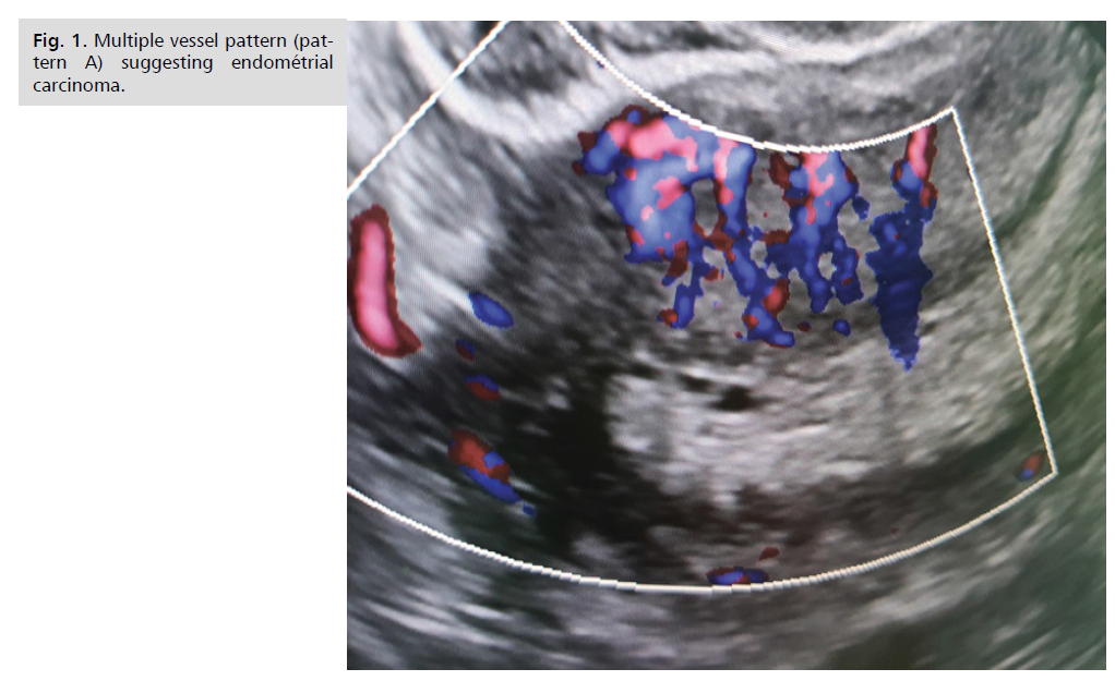
a) Multiple vessel pattern: Irregular branching corresponding to Endometrial carcinoma,
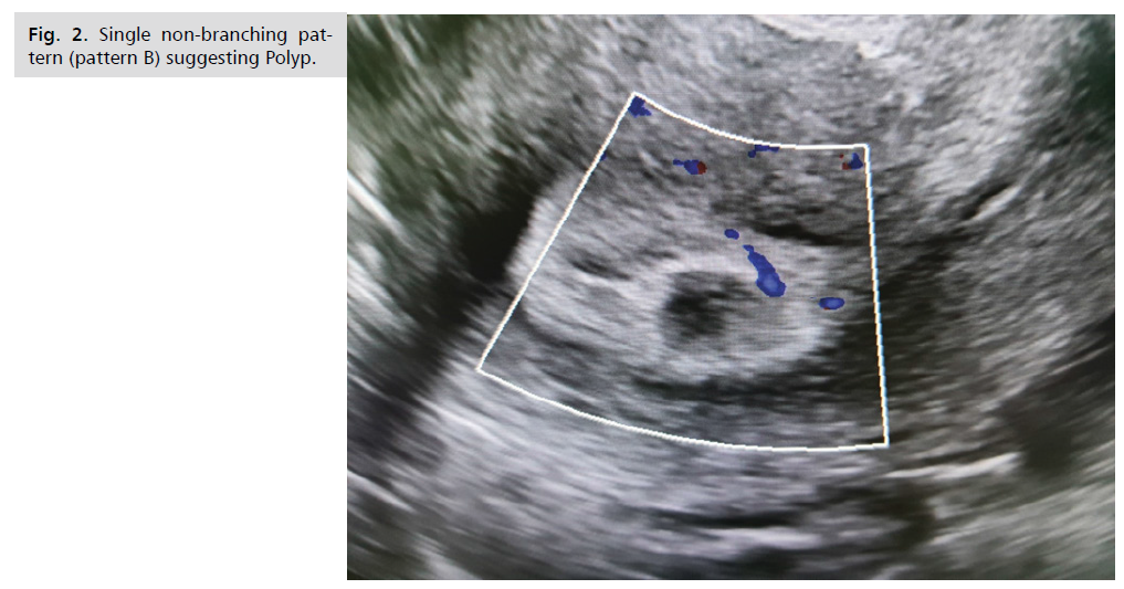
b) Single vessel pattern: With or without division as a character of polyp and
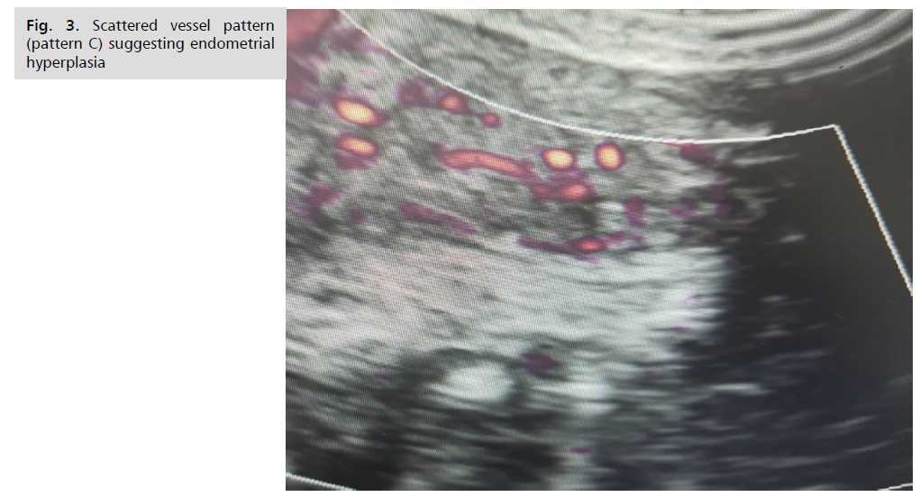
c) Scattered vessel pattern: Suggestive of hyperplasia [6].
The Spiral arteries were examined and flow velocity waveforms (FVW) were obtained to measure the resistant index (RI) and pulsatility index (PI).
Results
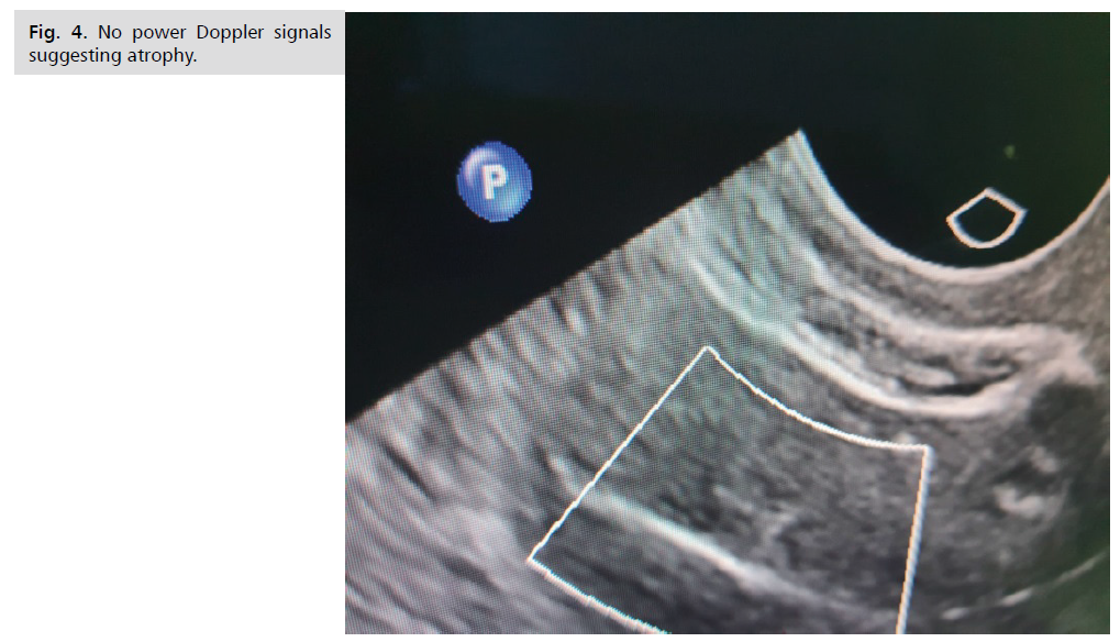
From November, 2019 to July, 2020, a group of 180 women complaining of post-menopausal bleeding were studied. The mean age was 59.25 ± 7.25 years (range 47-73), the mean body mass index was 31 ± 2.51 kg/m2 (range: 26-36). Transvaginal Power Doppler vascular patterns revealed 16 cases (8.9%) showed multiple branching vessels pattern (pattern A) (Fig. 1.) strongly correlated with endometrial cancer, 31 cases (17.2%) showed single non branching vessel (pattern B) (Fig. 2.) suggesting polyp and 61 cases (33.9%) showed scattered vessel pattern (pattern C) (Fig. 3.) suggestive of hyperplasia, and no power Doppler signals were detected in 72 cases (40%) (Tab. 1.) (Fig. 4.).

Fig 1. Multiple vessel pattern (pattern A) suggesting endométrial carcinoma.

Fig 2. Single non-branching pattern (pattern B) suggesting Polyp.

Fig 3. Scattered vessel pattern (pattern C) suggesting hyperplasia.

Fig 4. No power Doppler signals suggesting atrophy.
| Variables | Carcinoma (n=16) | Polyp (n=33) | Hyperplasia (n=63) | Atrophy (n=68) | χ2 | p | |
|---|---|---|---|---|---|---|---|
| Simple (n=48) | Atypical (n=15) | ||||||
| Pattern A (n=16) | 16 (100%) | 0 | 0 | 0 | 0 | 120 | <0.001 |
| Pattern B (n=31) | 0 | 31 (93.9%) | 0 | 0 | 0 | ||
| Pattern C (n=61) | 0 | 0 | 46 (95.8%) | 15 (100%) | 0 | ||
| no Doppler signal (n=72) | 0 | 2 (6.1%) | 2 (4.2%) | 0 | 68 (100%) | ||
Tab. 1. Comparison between histopathological diagnosis and different transvaginal power Doppler vascular patterns.
The histopathological diagnosis obtained via endometrial sampling was as follows: 16 cases (8.9 %) diagnosed as endometrial carcinoma, 33 cases (18.3%) had benign polyps, 63 cases (35%) had hyperplasia, 48 cases (26.6 %) of them had simple hyperplasia and 15 cases (8.3%) had hyperplasia with atypia and 68 cases (37.7 %) had thin endometrium with non-specific findings diagnosed as atrophy (Tab. 1.).
Taking histopathological biopsy as the gold standard, the diagnosis assigned using transvaginal power Doppler vascular pattern was confirmed in all cases of endometrial carcinoma but it missed the diagnosis in 4 cases; 2 with endometrial polyp and another 2 with endometrial hyperplasia. There was no false positive diagnosis using transvaginal power Doppler vascular patterns for any of the reported pathologies. The diagnostic performance of transvaginal power Doppler vascular patterns for detection of endometrial pathologies are shown (Tab. 2.) with 100% accuracy for diagnosing endometrial carcinoma, 94.12% for diagnosing endometrial polyp and 96.8% for diagnosing endometrial hyperplasia.
| Variables | Sensitivity | Specificity | Positive Predictive Value | Negative Predictive Value | Accuracy |
|---|---|---|---|---|---|
| Endometrial carcinoma | 100% | 100% | 100% | 100% | 100% |
| Polyp | 93.94% | 100% | 100% | 98.76% | 94.12% |
| Endometrial hyperplasia | 96.83% | 100% | 100% | 98.40% | 96.80% |
Tab. 2. Diagnostic performance of transvaginal power Doppler vascular patterns in diagnosis of different endometrial pathology.
The mean endometrial thickness and spiral arteries color Doppler indices are shown (Tab. 3.). Cases of endometrial carcinoma showed a highly statistical difference compared to other benign conditions. In patients with endometrial cancer spiral arteries PI was found to be significantly lower than the benign lesions (p <0.001). Spiral arteries RI was also lower in endometrial cancer cases compared to endometrial polyp, hyperplasia and atrophy (p <0.001).
| Variables | Carcinoma (n=16) | Polyp (n=33) | Hyperplasia (n=63) | Atrophy (n=68) | p |
|---|---|---|---|---|---|
| Endometrial thichness | 18 ± 6 | 10.6 ± 3.9 | 8.6 ± 4.2 | 3 ± 1.2 | <0.001 |
| Pulsatility Index | 0.71 ± 0.6 | 0.95 ± 0.14 | 0.93 ± 0.7 | 0.9 ± 0.5 | <0.001 |
| Resistance Index | 0.42 ± 0.05 | 0.9 ± 0.1 | 0.74 ± 0.19 | 0.95 ± 0.1 | <0.001 |
Tab. 3. Comparison between histopathological diagnosis and endometrial thickness as well as transvaginal color Doppler indices.
Discussion
This prospective study assessed the validity of transvaginal power Doppler vascular pattern as a diagnostic tool in cases presented with postmenopausal uterine bleeding in comparison to histopathological diagnosis from obtained via dilatation and curettage. In this study, we noticed that atrophy is the commonest pathological lesions as a cause of PMB. This is in agreement with previous studies which reported that the commonest cause of uterine bleeding in postmenopausal women is atrophy [7,8].
Our results indicated that the use of three vascular patterns depicted by power Doppler showed high sensitivity and specificity in diagnosing endometrial pathologies in postmenopausal bleeding.
All cases with endometrial cancer in this study showed multiple vessels pattern (pattern A) 16 of 16 cases (100%) with 100% sensitivity, specificity positive and negative predictive value. This result is in agreement with Dragojević et al., who stated that malignant lesions are associated with angiogenic alterations, bizarre atypical vascularization, increased intramural blood flow and increased flow in blood vessels of pelvic organs. They also stated that the presence of newly formed and multiple densely arranged myometrial and endometrial blood vessels is highly suggestive for malignancy [9].
This study showed that power Doppler vascular pattern 61 of 63 cases with endometrial polyp and no false positive diagnosis with 96.83%, 100%, 100%, 98.4% and 96.8% sensitivity, specificity, positive and negative predictive value and accuracy, respectively. Power Doppler in this study also diagnosed 31 of 33 cases (93.9%) with endometrial hyperplasia and there was no false positive cases, its sensitivity, specificity, positive and negative predictive value and accuracy was 93.94%, 100%, 100%, 98.76% and 94.12%, respectively. Contrary to our results, Alcazar et al., 2013 found the scattered vascular pattern of the hyperplasia in (8 of 15 cases 53%) and they concluded that power Doppler is not useful to detect endometrial hyperplasia due to low sensitivity [10]. This difference may be due to the difference in the sample size from the present study.
We also evaluated the vascular indices of the sub-endometrial vessels which showed correlation between benign and malignant endometrium, where both spiral artery RI and PI were significantly lower in endometrial carcinoma than other benign conditions (p <0.001). Malignant endometrium had the lowest RI compared to other histopathological diagnosis.
These results were in agreement with previously reported studies. Szpurek D et al., reported low vascular resistance index (RI =0.42 ± 0.05) with endometrial malignancy [11]. Kurjak and Kupesic reported RI higher in cases with endometrial hyperplasia (0.70) than endometrial carcinoma (0.60) [12]. Also, Amit et al., used power Doppler to identify endometrial vessels and used a cut off RI <1.0 as selection criterion to discriminate between endometrial carcinoma and benign lesions [2]. Against to this finding was the work of Opolskiene et al., which reported that flow indices could not discriminate well between benign and malignant endometrium [4].
This study confirmed that power Doppler study is useful in differentiating endometrial carcinoma from benign endometrial pathologies. Second, power Doppler is highly accurate in diagnosing endometrial carcinoma with accuracy reaching 100% in this study but this result should be interpreted with caution due to small size of cases (only 16 cases). Third spiral arteries Doppler indices highly correlate with endometrial carcinoma although we could not present a cut off value suggestive of malignancy.
Conclusion
Power Doppler ultrasound is a useful adjunct tool in diagnosis of endometrial pathology in cases with postmenopausal bleeding and shows good reliability in the discrimination between endometrial carcinoma from other benign endometrial conditions.
References
- Aboulfotouh M, Mosbeh MH, Elgebaly AF, et al. Transvaginal power Doppler sonography can discriminate between benign and malignant endometrial conditions in women with postmenopausal bleeding. Middle East Fert Soc J. 2012;17:22-29.
- Amit A, Weiner Z, Ganem N, et al. The diagnostic value of power Doppler measurements in the endometrium of women with postmenopausal bleeding. Gynecol Oncol. 2010;77(2):243-247.
- Kupesic S, Kurjak A. Uterine lesions. Medicinski Glasnik. 2009:49-59.
- Opolskiene G, Sladkevicius P, Valentin L. Ultrasound assessment of endometrial morphology and vascularity to predict endometrial malignancy in women with postmenopausal bleeding and sonographic endometrial thickness 4.5 mm. Ultrasound Obstet Gynecol. 2010;30(3):332-340.
- KabilKucur S, Temizkan O, Atis A, et al. Role of endometrial power doppler ultrasound using the international endometrial tumor analysis group classification in predicting intrauterine pathology. Arch Gynecol Obstet. 2013;288:649-654.
- Timmerman D, Bourne T, Valentin L, et al. Terms, definitions and measurements to describe the sonographic features of the endometrium and intrauterine lesions: A consensus opinion from the international endometrial tumor analysis (IETA) group. Ultrasound Obstet Gynecol. 2010;35:103-112.
- Litta P, Merlin F, Saccarid C. Role of hysteroscopy with endometrial biopsy to rule out endometrial cancer in postmenopausal women with abnormal uterine bleeding. Maturitas. 2005;50(2):117-123.
- John I, Gentry-Maharaj A, Burnell M, et al. Sensitivity of transvaginal ultrasound screening for endometrial cancer in postmenopausal women: A case–control study within the UKCTOCS cohort. Lancet Oncol. 2017;12(1):38-48.
- Dragojovic, Hiemstra E, Trimbos JB, et al. Power Doppler area in the diagnosis of endometrial cancer. Int J Gynecol Cancer. 2010;20(7):1160-1165.
- Alcazar JL, Castillo G, Minguez JA, et al. Endometrial blood flow mapping using transvaginal power Doppler sonography in women with postmenopausal bleeding and thickened endometrium Ultrasound Obstet Gynecol. 2013;21:583-588.
- Szpurek D, Sajdak S, Moszynski R, et al. Estimation of neovascularisation in hyperplasia and carcinoma of endometrium using a ‘power’ angio-Doppler technique. Eur J Gynaecol Oncol. 2010;21:405-407.
- Kurjak J, Kupesic H. Color Doppler ultra-sonographic features of uterine arteriovenous malformations: Report of two cases. Ultrasound Obstet Gynecol. 2019.
Author Info
Sally AR Kotb1*, Nahla M Awad2 and Amal S Zaghlaul12Department of Obstetrics and Gynecology, Early Cancer Detection and Gyn Endoscopy Unit, Maternity Hospital, Ain Shams University, Cairo, Egypt
Copyright:This is an open access article distributed under the terms of the Creative Commons Attribution License, which permits unrestricted use, distribution, and reproduction in any medium, provided the original work is properly cited.